digestive system of a lion
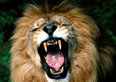
Lions have specialised teeth to suit their diet and lifestyle. The pre-molars and molars have evolved into the carnassial sheer which is effective at slicing through skin and muscle from the bone into swallowable chunks (Ewer, 1973).
50% of meat extraction is done through puling motions of the neck and 30% using paws (van Valkenburgh, 1996).
Being obligate carnoveres, lions have no requirement for carbohydrates in their diet, therefore do not produce salivary amylase within the buccal cavity (Herdt, 2002).
50% of meat extraction is done through puling motions of the neck and 30% using paws (van Valkenburgh, 1996).
Being obligate carnoveres, lions have no requirement for carbohydrates in their diet, therefore do not produce salivary amylase within the buccal cavity (Herdt, 2002).
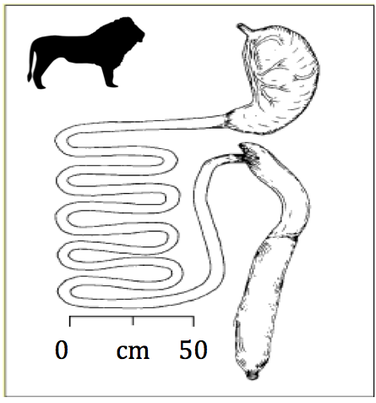
The lion oesophagus is approximately 70-80cm (Smith et al, 2006). Chunks of food are swallowed and travel through the oesophagus to the stomach by perilstalsis. The lion stomach is approximately 20% of their bodyweight (Smith et al, 2006). This is to enable storage of sizeable, infrequent meals. The average length between kills in the wild has been recorded as 1.5 - 3.5 days (Altman, 2005) but has been recorded to as much as 8 days in Kalahari lions (Eloff, 1984).
The stomach consists of four functionally distinct zones; the oesophageal region, where there is some bacterial growth but no glandular secretions. The cardiac region has alkaline mucus secreting glands, consisting of glycoproteins, which protects the stomach lining from being digested by the proteolytic enzymes and acid (Cheeke and Dierenfeld, 2010). The Fundus gland and pyloric regions are the sites of other gastric secetions, including hydrochloric acid (HCl). HCl provides a low pH within the stomach inorder to kill any bacteria consumed, and cause hydrolysis of proteins and polysaccharides alongside denaturation of proteins. The proenzyme pepsinogen is activated by the low pH, forming pepsin, to begin protein digestion. The broken down food exits via the pyloric region and enters the small intestine (Cheeke and Dierenfeld, 2010)
The entire length of the adult lion small intestine is 6 to 7 metres (Smith et al, 2005), comprising the duodenum, jejunum and ileum. The gall bladder secretes bile for emulsification of fats. Pancreatic enzymes, including trypsin, amylase and lipase, are released into the duodenum to aid digestion of large protein, carbohydrate and lipid molecules into amino acids, monosaccarides, fatty acids and glycerol ready for absorption (Stevens and Hume, 1995). Finger like villi cells, provide a large surface area for the absoprtion of essential nutrients, primarily within the jejunum. (Cheeke and Dierenfeld, 2010). Unlike herbivores, carnivores have no requirement for the caecum. In lions, it is approximately 10cm and positioned in reverse to that of the human tract. It is believed to have evolved this way to allow consumption of bone fragments and porcupine quills without blocking the tract. (Smith et al, 2006)
The large intestine of an adult lion is approximately 1 metre. Its main function is the absorption of water and electrolytes. The intestine of domestic cats maintains bacterial colonies comparable to herbivores. These protect against invading bacteria, stimulate gastrointestinal function such as motility and digest fibre sources to produce volatile fatty acids (Suchodolski, 2011). Recent research has revealed that carnivores have hind-gut fermentation of poorly enzymatically digestible tissues, such as fur and bones (Depauw et al, 2013), resulting in slow-release leptin and satiation for longer between kills.
The simple digestive tract of lions allows for efficient digestion of prey animals but, in contrast with herbivores, limited digestion of more complex fibre sources.
